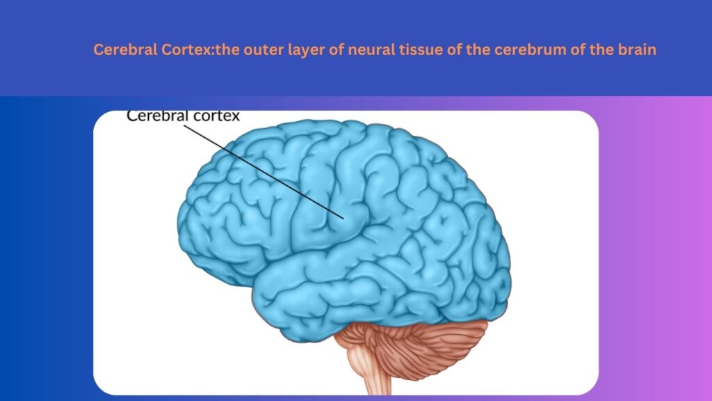
The mammalian brain’s cerebrum is surrounded by a covering of neuronal tissue called the cerebral cortex. It has nerve cells in up to six layers. It is frequently called “grey matter” and is protected by the meninges. The cortex is grey because the nerves there don’t have the insulation (myelin) that gives most of the rest of the brain its white appearance.
The Cerebral Cortex: What Is It?
The outermost portion of your brain is called the cerebral cortex. It has many folds on its surface, giving it a wrinkled appearance. The folds are made up of numerous sulci, or deep grooves, and gyri, or elevated areas. These folds increase the cerebral cortex’s surface area, which enables more nerve cells to process enormous amounts of information. About half of the overall volume of your brain is made up of the cerebral cortex. There are between 14 billion and 16 billion nerve cells in your cerebral cortex, comprising six nerve cell layers. It has a thickness between two millimeters (mm) and four mm (0.08 inch to 0.16 inch). The four lobes of your cortex are frontal, parietal, temporal, and occipital. Different kinds of information are processed by each of these lobes. The higher-order functions of the human brain, such as language, memory, reasoning, thought, learning, decision-making, emotion, intelligence, and personality, are collectively controlled by your cerebral cortex.
Why Is Grey Matter Referred To As The Cerebral Cortex?
Your brain’s outer layer of grey matter is made up of nerve cell bodies, including dendrites, the end of nerves. The portion of a nerve cell called dendrites is responsible for receiving chemical messages from neighbouring cells. Your cerebral cortex appears grey because it lacks myelin, a fatty substance that covers nerves. Bundles of axons, the long, myelin-wrapped centre section of a nerve cell, make up the white matter in your brain. The tissue is whitish because of the myelin.
What Distinguishes The Cerebral cortex from the cerebrum?
Your cerebral cortex, which is located on top of your cerebrum, is its outer layer. The greatest part of your brain is called your cerebrum. Your brain is split into two hemispheres by your cerebrum. The corpus callosum, a network of nerve fibres, connects the hemispheres together. Your two hemispheres may communicate with one another thanks to the corpus callosum.
What is the Neocortex?
The majority of your cerebral cortex is viewed as the neocortex. “Neo” signifies new. Your neocortex is so named on the grounds that its appearance is believed to be generally new in vertebrate advancement. In people, 90% of the cerebral cortex is the neocortex.
What Are The Cerebral Cortex’s Functions?
Many high-level processes, including logic, emotion, thought, memory, language, and awareness, are carried out by your cerebral cortex. Your brain has different functional areas connected with each lobe.
Functions Of The Frontal Lobe
Your frontal lobe is located behind your forehead in the front of your brain. Your frontal lobe has the following functions:
- Making decisions and resolving issues.
- thinking consciously.
- Attention.
- control over emotions and behaviour.
- production of speech.
- Personality.
- Intelligence.
- Body motion.
This lobe’s motor cortex, prefrontal cortex, and Broca’s region are noteworthy special areas. Your motor cortex is in charge of how you move. Thinking and problem-solving are examples of “executive functions,” which are controlled by your prefrontal cortex. Additionally, it guides and monitors other parts of your brain. Your frontal lobe’s Broca’s region plays a role in speech production.
What parts of the cerebral cortex are there?
Some researchers use a different approach to studying the brain and categorise the cerebral cortex’s regions according to their three primary types of functions: sensory, motor, and association areas.
Sensory areas:
- These parts of the cerebral cortex are where sensory data from the world and your senses is processed. Functions comprise:
- Iinterpreting visual data and recognising objects. The visual cortex is a part of your occipital lobe that processes these tasks.
- evaluating data from your body related to touch, temperature, location, vibration, pressure, and discomfort. The somatosensory cortex, a part of your parietal lobe, is responsible for processing these actions.
Motor areas:
These cerebral cortex regions are known as the “motor areas,” and they are involved in the voluntary contraction of muscles. Your frontal lobe processes these activities primarily.
Functions comprise:
Movement coordination between muscles.
Complicated movement planning.
using empathy and imitation to learn.
Association areas:
All four lobes are home to association areas, which connect and make processes more complex.
Functions comprise:
- Putting information from the sensory and motor systems into order and giving it meaning.
- Personality and emotional self-control.
- spatial reasoning and awareness.
- memory management.
- Think visually and remember things visually.
- Using language, music, and memory, create visual information.
What signs might indicate a damaged cerebral cortex?
The damaged region of the cerebral cortex determines the symptoms.
Experiencing frontal lobe damage
The following are signs that your frontal lobe has been hurt or damaged:
- Memory problems.
- Individuality varies.
- decision-making and problem-solving challenges.
- Attention deficits.
- behavioural changes, socially unacceptable behaviour, and emotional impairment.
- Aphasia is the inability to understand or use words.
- speech impairment (apraxia).
- flaccid hemiplegia is characterised by weakness, paralysis, and loss of muscle control on one side of the body.
- Dementia is another factor that can harm the frontal lobe.
Parietal lobe damage
- The following are signs that your parietal lobe is damaged:
- Making memories.
- Inability or difficulty writing (agraphia).
- problems with math.
- Numbness.
- Disorientation.
- uncoordinated hand-to-eye coordination.
- incapacity to recognise objects solely by touch (astereognosis).
- sensory loss.
- Aphasia.
- Apraxia.
A temporal lobe injury
The following are signs that your temporal lobe is damaged:
- Hearing problems.
- Memory problems.
- Recognising faces and objects is difficult.
- language disorders, including Wernicke’s aphasia, and communication problems.
- Epileptic seizures, developmental dyslexia, and Alzheimer’s disease are Further conditions that can harm the temporal lobe.
A occipital lobe injury
The following are signs that your occipital lobe is damaged:
- Having trouble focusing on multiple objects at once.
- Having trouble identifying things by sight.
- Blindness to colour.
- Vision-based hallucinations.
- Complete blindness.
FAQs
A- The outermost layer of the brain’s surface is called the cerebral cortex, and it is found on top of the cerebrum. The cerebral cortex is responsible for many mental and sensory functions, including memory, learning, reasoning, problem-solving, emotions, consciousness, and sensory perception.
A- The two cerebral hemispheres that make up the cerebrum are the cortex (sometimes known as grey matter) and the inner layer (also known as white matter). The frontal, parietal, temporal, and occipital lobes are the four lobes of the cortex. The primary focus of this review essay will be the functioning of the cerebral cortex.
A- The four lobes that make up the cerebral cortex are the frontal, parietal, temporal, and occipital lobes.
A- The forebrain, or prosencephalon The forebrain is the largest and most visible part of a mammal’s brain. The cerebral hemispheres are located in the cerebral cortex, which accounts for two-thirds of the brain’s total mass.
A- The nature of the cerebral cortex, its functions, and its location. Your cerebral cortex is in charge of all of the higher-order mental processes, including language, memory, reasoning, thought, learning, decision-making, emotion, intelligence, and personality.
Read Also:

